Electrophysiology
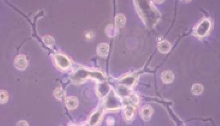
Electrophysiology studies the electrical properties of cells and tissues, enabling the characterization of electrical activity in excitable living cells (neurons, cardiomyocytes, etc..) and the understanding of nerve membrane ionic currents involved in molecular and cellular signaling.
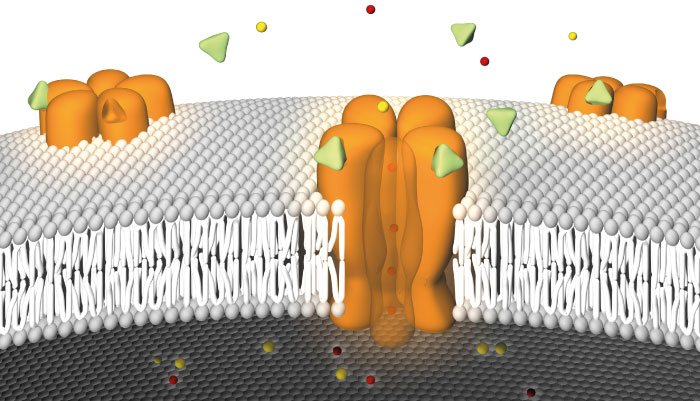
Located in cell, plasma and organelle membranes, ion channels play a crucial role in the modulation of physiopathlogical mechansisms in different cell type microenvironment; aberrant ion channel functioning is linked to several diseases and conditions affecting neuronal signaling, cardiac excitability and muscles activity.
Thanks to this peculiar role they represent the third most frequent biochemical class of targets for marketed small molecules behind enzymes and G-protein coupled receptors.
The electrophysiological study of ion channel functioning is thus a critical research task that could lead to advancement in therapeutics development and pathology mechanisms dissection.

ELECTRA Platform
Electrophysiological Assessment System
an innovative electrophysiological approach for intra and extracellular mechanisms investigation
ELECTRA AT A GLANCE
a review of the recording types and analysis methods provided with our platform
Extracellular Recordings
In extracellular recordings, an electrode is not inserted into a single cell, instead the electrodes are place in the extracellular fluid, near the cell of interest. Field potentials are probably the most common extracellular signals being recorded. Extracellular field potentials are local current sinks or sources that are generated by the collective activity of many cells. Usually, a field potential is generated by the simultaneous activation of many neurons by synaptic transmission.
Intracellular Recordings
Intracellular recording involves measuring voltage and/or current across the membrane of a cell. To make an intracellular recording, the tip of a fine (sharp) microelectrode must be inserted inside the cell, so that the membrane potential can be measured. Typically, the resting membrane potential of a healthy cell will be -60 to -80 mV, and during an action potential the membrane potential might reach +40 mVhe voltage measured by the electrode is compared to the voltage of a reference electrode, usually a silver chloride-coated silver wire in contact with the extracellular fluid around the cell.

Patch-Clamp
- The patch-clamp technique to study a single or multiple ion channels in cells.
- Current Clamp
- Voltage Clamp
Dose-Response Analysis
- Dose-response curves and an EC50 analysis thorough electrophysiological recording following during exposure to candidate molecules
Ion-channel Analysis
- Biophysical property characterization of current density, activation and inactivation curves of voltage-dependent ion channels.